Contrast Media
Contrast Media In Radiology
Written By Dr. Sherine Hamed
Specialist of Diagnostic Radiology – Teleradiology Medical Administrator

– Contrast media is a group of chemical agents developed to aid in the characterization of pathology by improving the contrast resolution of an imaging modality. Specific contrast media have been developed for every structural imaging modality and every conceivable route of administration.
– Contrast media (CM) is used in imaging techniques to enhance the differences between body tissues in images. The ideal contrast medium should achieve a very high concentration in the tissues without producing any adverse effects.
– In the last decades procedures employing CM have rapidly increased. Significant improvements in the composition of CM during the past few decades have made them safer and better tolerated.
– Iodinated and gadolinium-based contrast media are used daily in most radiology practices. These agents often are essential to providing accurate diagnoses and are nearly always safe and effective when administered correctly.
Types of contrast media:
1. Barium sulfate contrast media have been used for many decades, and are well-established as oral agents for the examination of the GI tract. Their use is generally restricted to radiographic and fluoroscopic examinations. Occasionally they are also used for CT examination of the GI tract (e.g. CT colonography in iodinated contrast media-allergic patients). They are cheap and well-tolerated by most patients, complications from their use are rare.
2. Iodinated contrast media are the mainstay contrast agents for radiographic, fluoroscopic, angiographic, and CT imaging. They are a versatile group of agents used for intravenous, oral, and other routes of administration, such as urethral and intra-articular.
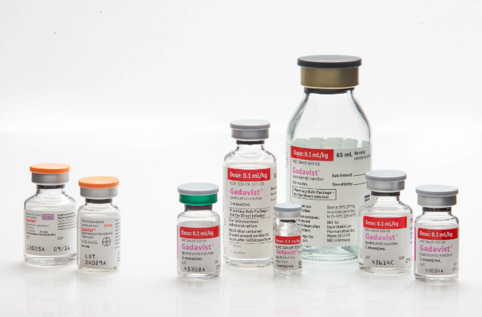
3. MRI contrast media are most commonly gadolinium-based contrast agents (GBCAs), which are the agents used for the vast majority of contrast-enhanced MRI scans.
4. Ultrasound contrast media have been gaining traction in recent years. e.g. characterization of liver lesions and sonohysterography.
Routes of administration:
There are multiple different routes by which substances can be transferred into the human body depending on the type of imaging modality that will be performed, the patient’s complaint, and the patient’s medical history or known pathology.
- Non-invasive:
The most common non-invasive method is per oral (PO): the most common route for medication. Blood vessels, gastrointestinal (GI) tract, organs, and other body tissue that temporarily contain the iodine-based or barium compounds change their appearance on x-ray or CT images.
Invasive:
The most common invasive methods are: Iodine-based contrast materials injected into a vein or artery (intravenous or intra-arterial) are used to enhance X-ray (including fluoroscopic images) and CT images. Iodine-based contrast materials are also commonly injected into the arteries during angiographic procedures. Gadolinium injected into a vein (intravenously) is used to enhance MR images.
Precautions:
- Propper history recording of symptoms, list of surgeries of the area being scanned, list of medications, and list of allergies.
- Old studies that have been done before the current study.
- Previous history of administration of contrast media, and history of developing adverse reactions with the date registered for that.
- Medications are currently used for chronic diseases.
- For females: The date of last menstrual period, current pregnancy, and breastfeeding.
Adverse reactions:
Mild:
Signs and symptoms: are self-limited without evidence of progression Nausea, vomiting Altered taste Sweats Cough Itching Rash, hives Warmth (heat) Pallor Nasal stuffiness Headache Flushing Swelling: in eyes, face Dizziness Chills Anxiety Shaking
Treatment: Observation and reassurance. Usually, no intervention or medication is required; however, these reactions may progress into a more severe category.
Moderate:
Signs and symptoms: Due to reactions that require treatment but are not immediately life-threatening like Tachycardia/bradycardia Hypotension Bronchospasm, wheezing Hypertension Dyspnea Laryngeal edema Pronounced cutaneous Pulmonary edema reaction.
Treatment: Prompt treatment with close observation.
Severe:
Signs and symptoms: Life-threatening with more severe signs or symptoms including Laryngeal edema Profound hypotension Unresponsiveness (severe or progressive) Convulsions Cardiopulmonary arrest Clinically manifest arrhythmias
Treatment: Immediate treatment. Usually requires hospitalization.
MRI contrast agents:
Gadolinium-based contrast agents can be used safely for imaging patients in most settings. The risk of allergic reaction is less than with CT agents, and MR contrast agents are not nephrotoxic. However, extremely ill inpatients with renal insufficiency may be a risk for a rare systemic disease like nephrogenic systemic fibrosis (NSF). Carefully considering the risks and benefits of a contrast-enhanced MR examination can minimize the possibility of developing NSF in high-risk inpatients.
Conclusion:
Although contrast agents are widely used with safe outcomes and little or no side effects, adverse reactions nonetheless may occur. They may be severe, and they may progress rapidly. Successful patient management during contrast-enhanced examinations requires all of the following:
- Knowledge of the patient’s medical history.
- Patient preparation, including premedication, if necessary.
- Proper selection of the agent to be used.
- Knowledge of the pathophysiology of contrast reactions.
- Prompt recognition and accurate assessment of reactions.
- Immediate availability of necessary equipment and drugs.
- Adequate prior planning and training.
- Current knowledge of medications and other treatment options.
On Rology platform, our mission is to provide radiology reporting with high-quality and accurate details. For more information about our teleradiology platform, contact us via email info@rology.net or visit our website www.rology.health

